Why assessing the aneurysm wall stability – and not the rupture risk – to counsel well
Webinar von Prof. I. Wanke und Prof. D. Rüfenacht

Why assessing the aneurysm
The intracranial aneurysm disease
The intracranial aneurysm disease exhibits with a significant prevalence of approx. 3% a rather small overall incidence of rupture, with less than one out of 300 aneurysms per year only. In view of these numbers and to avoid over-treating the increasingly incidentally discovered, asymptomatic patients with an aneurysm, there is need to refine currently practiced clinical methods of identifying patients, that warrant endovascular or surgical treatment. Further, with current treatment indication based on size mainly, and with increasing incidental findings in a size range similar to small ruptured lesions, there is room to look for better, potentially more reliable biomarkers than size only.
Parallel to the increasing power of identifying intracranial aneurysms incidentally with currently available medical imaging practice, the understanding of disease biology has rapidly evolved and enriches current knowledge on how aneurysms are formed, why they grow and what may lead to their rupture. This recent progress in disease understanding has brought biological processes of wall degenerative remodeling involving inflammation in the focus of aneurysm assessment. When it comes to counsel patients and decide together with them, on how to proceed particularly in front of a small, i.e. less than 10 mm sized incidental aneurysm finding, and in view of made progress in disease understanding, clinicians as us have looked for a re-orientation. Our current clinical practice has moved forward from the standard “evidence-based medicine method” to embrace these view-points enriched with recent knowledge, that put the aneurysm wall at center stage. Nowadays, for a personalized aneurysm wall assessment, we attempt to assess and are investigating a patient’s individual aneurysm wall condition in addition to the recognized parameters. This further requires an understanding and measure of wall stability criteria allowing for identifying critical differences, that will lead to an individual decision to either observe only or that would argue rather in favor of an active, potentially dangerous, invasive treatment by endovascular or surgical means. In summary, we wish to differentiate a stable from an instable lesion status.
Therefore, with a search for instability, signs of inflammatory aneurysm wall degenerative remodeling are at the center of current investigations and our efforts therefore focus on aneurysm wall imaging. Further, with mechano- biological concepts recognizing the role of local blood flow conditions when it comes to physical forces that govern the process, flow assessment and shear stress calculations are of interest as well. Therefore, advanced CFD knowledge has identified a local “hostile hemodynamic environment” as a critical influencing and risk factor to consider. Whether, biological assessment via markers will play an additional role, such as could be harvested in samples of the blood or the CSF, the future will show. In our clinical setting, investigating the aneurysm wall for signs of instability, MRI is efficient and best capable of showing the vessel wall, the vascular lumen morphology and the blood flow, all in one examination. In our opinion, MRI based image information provides most efficiently imaging biomarkers representing the aneurysm wall status, where we may deduct an actual status of stability or instability. Specific MRI/MRA sequences may include information such as blood flow, thrombus formation, lumen size and vascular geometry, parent artery geometry and wall enhancement (Gadolinium), and potential interaction with a given perianeurysmal environment. This information allows for evaluating efficiently wall stability using following criteria:
- No growth / small size of an aneurysm over time (stable) versus
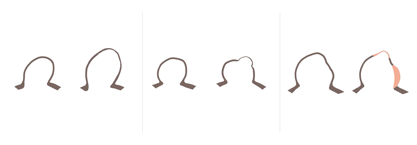
growing / large size (instable). - Regular, round shape (stable) versus irregular shape, implying
irregular wall thickness and lesser biomechanical strength
(instable). Irregularity has been shown to be associated with an
unstable, evolving clinical course. - Non-enhancing wall (stable) versus enhancing wall segments
(instable). Permeability of the wall for Gadolinium contrast material
indicates loss of endothelial function. This likely occurs due to
damaged endothelium (areas exposed to HWSS?) or due to
presence of slow flow, clot overlaying endothelium (areas exposed
to LWSS?), or a combination of both. Enhancing lesion are thought
to indicate areas of inflammation, consequently softening of the
aneurysm wall.
In addition to such “cumulative” criteria of instability, we consider location (“hostile hemodynamic stress”?), and other recognized risk factors, such as family history of SAH, history of smoking, hypertension and others. Presence of multiple signs of instability brings forward the decision to move forward rapidly with treatment.
Using these criteria for identifying an “aneurysm stability status”, we enroll about 50% of incidental findings for treatment and do observe over time a change of stable lesions towards instability in about 5-10% only.
In case a lesion is considered stable, we perform repeat MRI assessments at 6mths, 12mths and more. With aneurysm disease considerd to be a chronic disease, patients having undergone treatment will get a similar follow-up.

Prof. Dr. med.
Isabel Wanke